Anatomy of the Hip
Bones
Your hip joint is a ball and socket joint.
- Femur: The ball is made up of the top of the thigh bone or femur.
- Pelvic bone: The socket is the cup shaped bone that is part of the pelvis. This is called the acetabulum.
Trochanter: This is part of the upper femur. There is a large outer bony prominence called the Greater Trochanter and a smaller one inside the groin called the Lesser Trochanter. The part that you can feel as the outermost bit of the hip that you lie on is the Greater Trochanter.
Function of the Hip joint
Your hip is essential for normal everyday activity and takes 100% to 50% of your body weight when walking. This can multiply to several times body weight when doing sports and running activity.
Articular Cartilage (Hip): The articular cartilage covers the head of the ball and inside of the socket to absorb stress and allow smooth joint movement with minimal friction.
Labrum: This is a lipped cartilage ring on the outside of the socket that forms a seal for the ball and socket joint. The fluid inside the joint helps with smooth, pain-free movement.
Core Muscles of the Hip
The hip joint is supported by several core muscle groups:
1.Gluteal Muscles: These are the buttock muscles that help stabilise the pelvis for efficient walking and running activity. They assist in backward (extension), out to the side (abduction), and outward rotation (external rotation) movements of the leg. They are three in total:
2.Gluteus Maximus: The largest muscle in the body. This forms the outermost muscle layer and attaches the pelvis to the upper thighbone. It helps maintain balance as you walk or run. As the quadriceps muscles move the leg forward, the gluteus maximus contracts to propel the body forwards with force. This also lifts you from a squatted position and when climbing stairs.
3.Gluteus Medius: This lies underneath the Maximus layer and covers the outer upper thigh bone (Greater Trochanter) with a cuff of muscle that allows the leg to lift out to the side and is important for normal walking or running. This helps to swing your leg backwards and forwards when walking or running. Weakness in this muscle is very commonly seen and results in abnormal walking/running patterns, which are inefficient and cause pain and fatigue.
Gluteus Minimus: This is the smallest muscle and lies directly over the outer half of the ball and socket joint.
Hamstrings Muscles: These three muscles attach the bony part of the pelvis that you sit on, to the back of the knee. They move the hip backwards in extension and flex the knee joint.
Hip Flexors: These muscles help to flex the thighbone forward onto the pelvis.
Psoas Muscle: This is one of the deepest core muscles arising from the inside of the pelvis and attaching to the inner thigh at the Lesser Trochanter. It is a major hip flexor and can cause pain and a snapping hip from over-exertion inflammation when an imbalance has occurred.
Quadriceps Muscle: This is a group four muscles that attach the pelvis to the patella bone and helps flex the hip and extend the knee joint.
Adductor Muscles: These pull the leg back in towards the body midline.
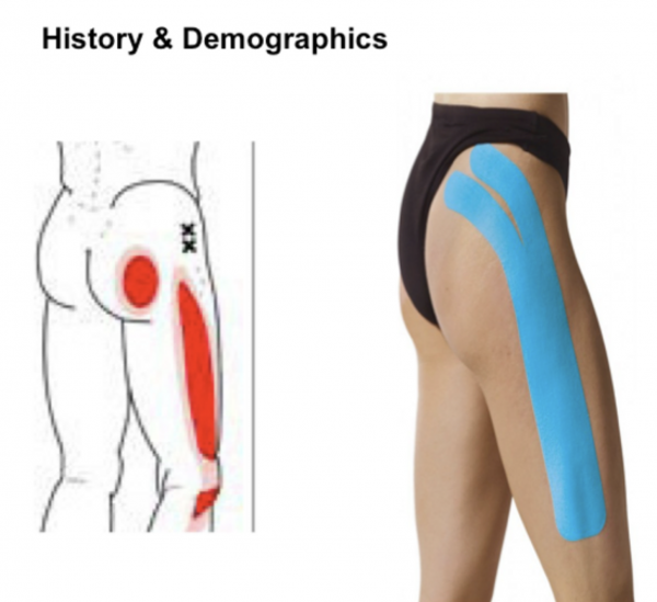
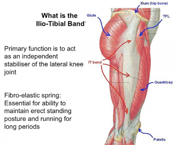
ITB (Ilio-Tibial band): This is a tight band of fibrous tissue that acts like a coiled spring storing energy that helps humans in their unique ability to stand, walk and run longer distances. It sits on the outer part of the pelvis, lies over the bony upper thigh and then inserts just below the outer knee joint. The tension and backwards/forwards balance position of this spring is delicately tuned by the core muscles and can cause symptoms of pain over the trochanteric area mimicking Trochanteric Bursitis or lower down over the knee joint causing ITB friction Syndrome or Runner’s Knee.
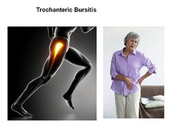
Trochanteric Bursa: Any area in the body that has skin overlying a bony prominence will have a thin two layer bag of fluid ( called a Bursa) to help lubricate the movement of skin over that prominence. It also helps cushion against a fall or lying on that hard part.
Sometimes that fluid filled bag becomes very agitated or inflamed causing Trochanteric Bursitis.


Surrey Orthopaedic Clinic Hip Specialist:


